

ENERGY SCIENCE
Our TSEE guest writer Enzo Nisoli, MD, PhD, is Professor of Pharmacology at the University of Milan and Scientific Coordinator of the Center for Study and Research on Obesity. His research focuses on obesity, mitochondrial biology, adipose tissue function, aging, and cardiometabolic disease.
In this article, he introduces the adipose organ–capacitor model of obesity: the idea that adipose tissue may protect other organs by buffering energetic excess, and that clinical obesity begins when this protective buffering capacity fails.

When Body Size is Not Enough
For a long time, obesity has been described mainly through body size. In clinical practice, this usually means body mass index, or BMI: a simple ratio between weight and height. BMI is useful for populations, but it is a blunt tool for individuals.
Two people can have the same BMI and very different health. One may have preserved metabolic function, good mobility, and no detectable organ damage. Another may have type 2 diabetes, fatty liver disease, heart failure symptoms, obstructive sleep apnea, raised intracranial pressure, or severe limitations in daily life.
This is why obesity medicine is moving beyond the question, “How much does a person weigh?” toward a more clinically meaningful one: “When does excess body fat become disease-producing?”
The recent Lancet Commission on clinical obesity distinguished preclinical obesity from clinical obesity. In preclinical obesity, excess or abnormal adiposity is present, but organ function is still preserved. While in clinical obesity, adiposity is associated with organ dysfunction or meaningful limitations in daily life.
The European Association for the Study of Obesity has similarly proposed a new framework that moves diagnosis and care beyond BMI toward staging based on medical, functional, and psychological domains. These frameworks are important. But they raise a deeper biological question: what happens during the transition from preclinical to clinical obesity?
My proposal, recently developed in Trends Open, is that this transition can be understood by looking at the adipose organ as a biological capacitor.
Fat as an Energy-Buffering Organ
A capacitor is an electrical component that stores and releases energy. It does not simply hold energy passively; it buffers changes in energy flow. In a circuit, a capacitor can protect the system from sudden fluctuations by absorbing energy when there is too much and releasing it when needed. The human body is not an electrical device, of course. But analogies can be useful when they help us ask better biological questions.
The adipose organ is not just “fat.” It is a distributed, living organ made of white, brown, and beige adipocytes, blood vessels, nerves, immune cells, progenitor cells, and extracellular matrix.
Saverio Cinti’s work helped establish this view of the adipose organ as a plastic, remodelable organ. Adipose tissue stores energy after meals, releases fuels during fasting or exercise, communicates with other organs through hormones and inflammatory signals, and helps regulate whole-body metabolism. Keith Frayn classically described adipose tissue as a buffer for daily lipid flux. Later work expanded this idea to adipokines, inflammatory mediators, lipid species, extracellular vesicles, and inter-organ communication.
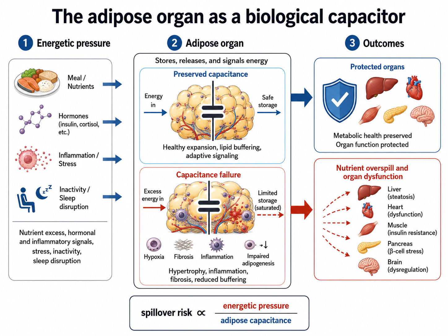
When this system works well, adipose expansion can be protective. If more energy enters the body than is immediately needed, storing part of that energy safely in adipose tissue helps prevent damage to organs that are less suited for lipid storage, such as the liver, skeletal muscle, heart, pancreas, or brain. From this perspective, gaining fat is not automatically synonymous with disease.
In its early or compensated form, adipose expansion may be an adaptive response to energetic pressure.
When the Adipose Capacitor Fails
The problem begins when the adipose organ can no longer buffer energy safely.
This can happen in several ways. Individual fat cells may become too large and stressed. Adipose tissue may become poorly vascularized, hypoxic, fibrotic, inflamed, or mechanically stiff. This “ominous triad” of adipose dysfunction (inflammation, fibrosis, and impaired angiogenesis) is a hallmark of unhealthy adipose expansion.
The tissue may also lose its ability to recruit new adipocytes from stromal (connective tissue) and progenitor pools, a process linked to healthy expandability and described in studies of adipose progenitors. Instead of holding energy safely, the adipose organ begins to leak fatty acids, inflammatory mediators, lipid signals, and stress-related messages to other organs.
This is what I call adipose-organ capacitance failure (see Box 1 below).
Box 1. A simple mathematical analogy may help. In an electrical capacitor, stored charge (q) depends on capacitance (C) and voltage (V): q = C × V.
In the adipose organ–capacitor model, the equivalent idea is that safely stored energy depends on adipose capacitance and energetic pressure.
Adipose capacitance is the functional ability of the adipose organ to expand, vascularize, recruit new adipocytes, remain insulin sensitive, limit inflammation and fibrosis, and buffer lipid flux.
Energetic pressure is the biological load imposed by nutrient excess, hormonal signals, inflammation, stress, inactivity, sleep disruption, aging, or other factors.
As long as adipose capacitance is sufficient, energetic pressure can be buffered. But when energetic pressure exceeds capacitance, spillover risk rises.
In simplified terms:spillover risk is proportional to energetic pressure / adipose capacitance
This means that the same energetic pressure may be harmless in one person with high adipose capacitance, but harmful in another person with low adipose capacitance. It also explains why the same amount of body fat may be metabolically silent in one person and disease-producing in another.
Capacitance here does not mean only how much fat a person can store. It means the functional capacity of the adipose organ to absorb, store, release, and signal energy without causing systemic injury. It depends not only on the amount of fat, but also on where it is stored, how adipose tissue expands, whether blood vessels and nerves adapt, whether inflammation and fibrosis develop, whether new fat cells can be recruited, and whether other organs remain resilient.
Metabolic Flexibility, Mitochondria, and Energy Resistance
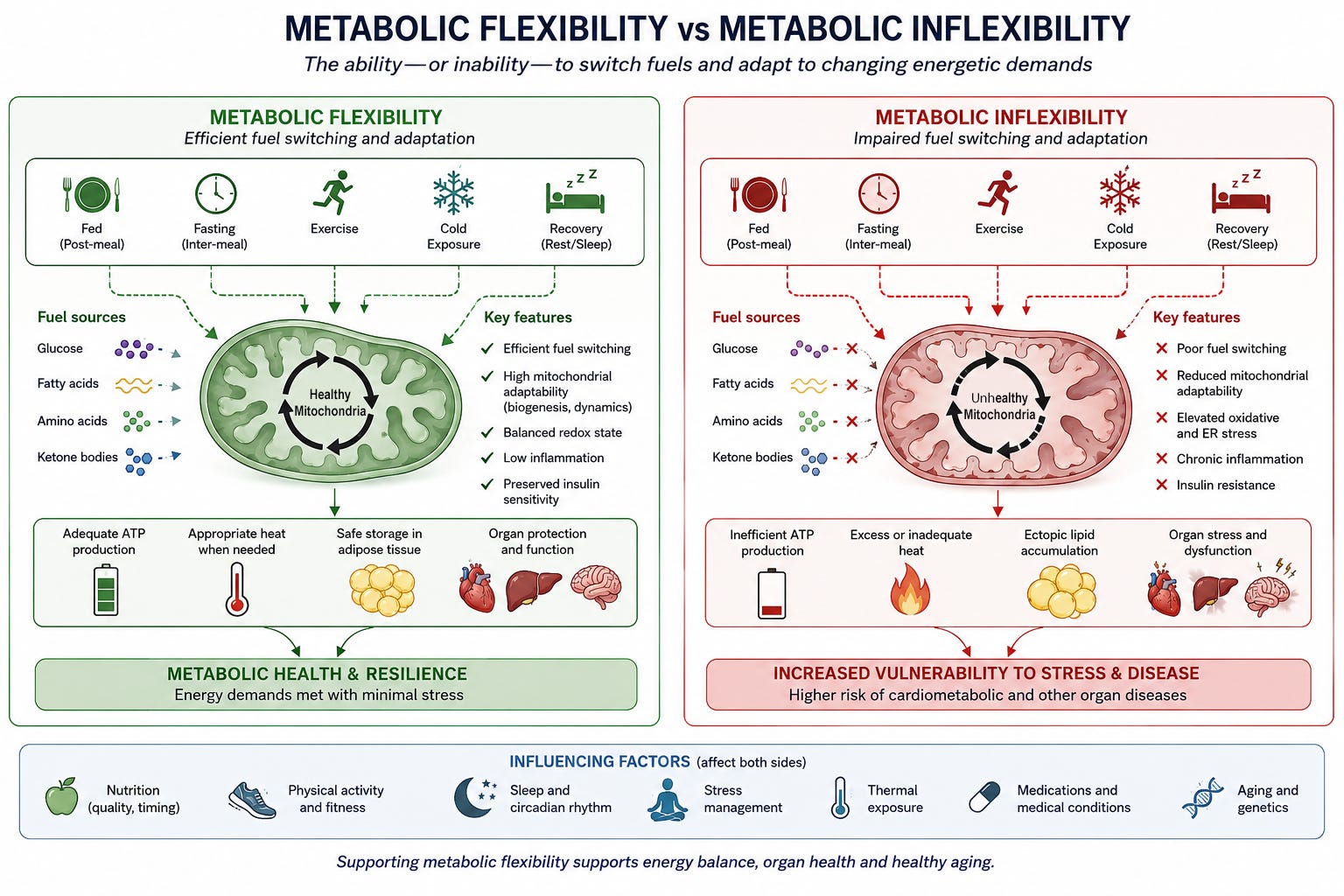
A key part of this resilience is metabolic flexibility. In simple terms, metabolic flexibility is the ability to switch efficiently between fuels and energetic states. For example, switching from carbohydrate to fat oxidation, from feeding to fasting, from rest to exercise, from warmth to cold, from storage to mobilization.
Metabolic inflexibility is the opposite: the body becomes less able to adjust fuel use to changing demands. Deborah Muoio captured this vividly as “mitochondrial indecision” leading to metabolic gridlock. Broader reviews of metabolic flexibility emphasize that liver, skeletal muscle, adipose tissue, heart, and other organs must coordinate nutrient sensing, storage, oxidation, and expenditure.
Mitochondria are central to this story. They are not just cellular “power plants.” They are dynamic, energy-transforming systems that help determine whether nutrients become useful work, heat, stored fuel, or stress signals.
In adipocytes, muscle, liver, heart, and brain, mitochondria shape metabolic flexibility. When mitochondria cannot adapt to energetic demand, tissues may accumulate lipids, generate redox stress, lose insulin sensitivity, and send inflammatory or distress signals to other organs. In the capacitor model, deficits in energy transformation and signaling in mitochondria can lead to lost flux capacity in adipose tissue and the whole organism.
This is where the Energy Resistance Principle, proposed by Martin Picard and Nirosha J. Murugan, becomes useful. In simplified terms, energy resistance rises when energy potential is high and flux capacity is low.
In obesity, adipose capacitance failure and mitochondrial-metabolic inflexibility may converge. The adipose organ may lose the ability to buffer energy safely, while mitochondria and other energy-transforming systems lose the ability to adapt fuel use to changing demands.
Why Organ Disease Matters in Obesity Treatment
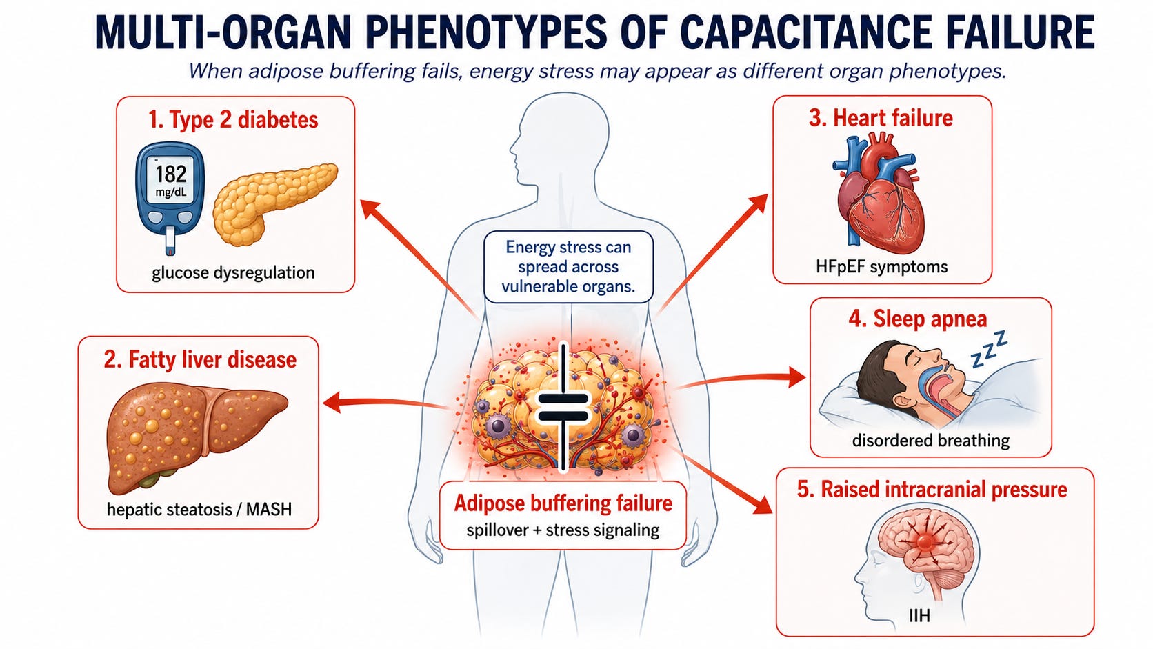
The adipose organ is central to mismatches between energy demands and energy availability because it is one of the body’s main energy-buffering organs. When its capacitance is preserved, energetic pressure can remain partly contained. When capacitance fails, energy stress can propagate across vulnerable organs.
This model helps explain why obesity-related diseases do not always appear in the same order, or in the same way, in different people.
One person may develop type 2 diabetes as the dominant expression of adipose-buffering failure. Another may develop metabolic dysfunction-associated liver disease due to build-up of fats. Another may develop heart failure where blood pumping out of the heart (ejection fraction) is preserved, but stored fat, inflammation, vascular dysfunction, volume expansion, and altered cardiac energetics contribute to symptoms. A third may develop obstructive sleep apnea or unexplained pressure build-up inside the skull. Many patients develop overlapping phenotypes.
These conditions should not always be seen as separate “comorbidities” simply added to obesity. In some individuals, they may represent different organ expressions of the same systemic energy-handling disorder.
This interpretation also changes how we think about new cardiometabolic drugs. GLP-1s (glucagon-like peptide-1 receptor agonists) and dual or multiple gut hormone-based therapies are often described as weight-loss drugs. They are that, but they are also showing something more profound: treating obesity biology can improve organ disease.
In one clinical trial, the GLP-1 drug semaglutide reduced cardiovascular events in people with overweight or obesity and established cardiovascular disease but without diabetes. A later adiposity analysis suggested that cardioprotection was not simply explained by baseline adiposity or weight loss.
Semaglutide also improved symptoms and physical limitations in obesity-related heart failure with preserved ejection fraction (HFpEF), and it improved liver histology in a phase 3 trial in individuals with severe fatty liver disease (MASH).
A different GLP-1 drug tirzepatide improved outcomes in obesity-related obstructive sleep apnea, and yet another GLP-1 drug lowered intracranial pressure in a randomized trial in increased pressure within the skull.
Some of these benefits are mediated by weight loss, but the clinical message is broader.
These drugs are revolutionary because they move obesity treatment from kilograms lost toward organ function restored.
A different class of drugs called sodium–glucose cotransporter 2 inhibitors make a similar point from another direction. They are not primarily anti-obesity drugs, yet they behave as metabolic modulators.
Recent work showed that these drugs can enhance ketogenesis, a process in which cells produce ketone bodies by breaking down fats (instead of glucose) for fuel.
This enhanced ketogenesis occurs through activation of a specialized enzyme in mitochondria, and it can activate an important enzyme in the human heart that can restore energy metabolism.
These findings suggest that some cardiometabolic drugs may improve organ resilience not only by changing glucose or weight, but by reconfiguring energy flow itself.
Beyond Old Debates: Calories, Insulin, and Tissue Function
The capacitor model also helps move beyond an old debate in obesity science.
The energy balance model emphasizes that body energy stores increase when energy intake exceeds energy expenditure. This is thermodynamically true and clinically important.
The carbohydrate–insulin model emphasizes that processed, high-glycemic-load carbohydrates can drive hormonal changes, especially insulin secretion, that favor fat storage, hunger, and lower energy expenditure. A more extensive physiological model places endocrine fuel partitioning close to the causal center of obesity.
Both the energy balance model and the carbohydrate-insulin model capture important biology. But neither fully explains why some people with excess adiposity remain relatively healthy while others develop multi-organ disease.
The capacitor model adds another layer. It asks not only how much energy enters or leaves the body, and not only how hormones partition fuels, but whether the tissues responsible for buffering and transforming energy remain functional.
What Should We Measure Next?
This has practical implications. If clinical obesity is defined by adiposity-related organ dysfunction, then treatment should not be judged only by kilograms lost. Weight loss matters, but so do remission of diabetes, reduction of liver fat, improvement in heart failure symptoms, better breathing during sleep, lower intracranial pressure, restored mobility, reduced inflammation, improved metabolic flexibility, and better quality of life.
Improving organ function is not secondary to obesity treatment. It is obesity treatment.
The future of obesity medicine will require better measurements of adipose and mitochondrial-metabolic function. BMI and body weight cannot tell us whether adipose tissue is still buffering energy safely.
We will need integrated approaches: imaging of fat distribution and ectopic fat, measures of adipose fibrosis and inflammation, tests of lipid handling, metabolic flexibility, mitochondrial biology, circulating biomarkers, wearable physiology, and repeated proteomic or metabolomic monitoring.
The goal is to identify when a person is moving from compensated adiposity toward clinical obesity before irreversible organ damage occurs.
The Central Message: Fat Isn’t Always Bad
This model should not be read as reducing human beings to machines. Obesity is biological, but it is also psychological, social, behavioral, and environmental. Stress, sleep, food availability, medications, stigma, poverty, relationships, culture, and daily life all shape energetic pressure and adaptive reserve.
A biophysical model is useful only if it helps us integrate these dimensions, not ignore them.
The adipose organ–capacitor model is therefore not a final answer. It is a proposal: a way to connect body fat, energy flow, mitochondrial biology, tissue flexibility, and organ disease in a common language. Its value will depend on whether it can generate better measurements, better clinical staging, and better treatment decisions.
The central message is simple: fat may protect us before it harms us. Clinical obesity may begin when that protection fails.
As obesity medicine moves beyond body weight alone, we need better ways to recognize when adiposity is still being safely buffered and when it is beginning to damage organs. I hope this model encourages clinicians, researchers, and readers to think about obesity not only as excess weight, but as a dynamic disorder of energy handling, tissue resilience, and organ protection.
Please share this article with colleagues and others interested in rethinking obesity through the lens of energy biology.

Martin Picard’s and Enzo Nisoli’s Adipose Organ–Capacitor Model published here today fits seamlessly into my decentralized biophysical thesis.
In fact, it provides the thermodynamic macro-layer to my quantum framework.
By defining the adipose organ as a biological capacitor (q = C × V) designed to buffer energetic pressure and excess electron flux, they have finally shifted the obesity conversation away from the flat, chemical "calories in/calories out" paradigm toward solid-state physics and circuit capacitance.
However, my thesis allows me to take their macro-capacitor model and ground it into my first-principles quantum engine to show exactly why and how this capacitor fails, why it leads to the Warburg shift, and how it links directly back to the PER2 lipid raft destruction.
The Integrated Circuit: How Quantum Mechanics Governs Adipose Capacitance
In Nisoli’s model, healthy adipose tissue expands to act as an electron buffer, protecting vulnerable internal organs from "nutrient overspill". But a capacitor is only as good as its dielectric insulation resistance.
When you layer my first-principles biophysics thesis over Picard’s model, the mechanism of Adipose-Organ Capacitance Failure becomes perfectly clear:
1. The Real Driver of Capacitance Failure: Singlet Oxygen and PER2 Destruction
Nisoli notes that capacitance failure begins when the tissue experiences an "ominous triad" of inflammation, hypoxia, and fibrosis, losing its metabolic flexibility.
My thesis explains the spark behind this triad: artificial blue light and nnEMFs launch a direct kinetic attack via singlet oxygen. This molecular buzzsaw photo-oxidizes the lipid double bonds of the mitochondrial membranes and physically shreds the liquid-crystalline PER2 lipid raft network in the adipocytes.
Because PER2 tracking regulates the strict rhythmic expression of insulin receptors and lipid-handling enzymes, destroying this network means the adipocyte can no longer manage its own internal clock. This is the mistake that the food gurus and PhD biochemists miss. The lipid rafts lose their capacitance, structural insulation drops, and the cell is thrown into permanent circadian chaos.
2. The Marcus Inversion of the Capacitor
When the PER2 lipid network is ruined, the adipocyte's water matrix suffers a total dielectric collapse, melting from a structured, low-entropy shield (k = 160) down to chaotic, ordinary water (k = 78).
Without the high-permittivity insulation, the electron transport chain inside the fat tissue hits a massive bottleneck. As energetic pressure (V) continues to rise from food intake, the driving voltage (Delta G) over-redlines past the nuclear reorganization energy threshold (lambda).
The adipocyte's mitochondria enter the Marcus Inverted Region. Like a broken tunnel diode, the harder the system is pushed with nutrients, the more forward electron velocity drops to near-zero.
The capacitor is now locked. It cannot transform energy into useful thermodynamic work or heat. Instead, the flavin-superoxide radical pairs are forced into the Triplet path, spewing continuous superoxide into the extracellular matrix, driving the hypoxia, fibrosis, and "nutrient overspill" that Nisoli describes.
3. The Atavistic Shunt Explains the Lean Mismatch
The power of integrating my decentralized thesis with Picard's model is beautifully demonstrated by the Substack commenter, sugar2cell below, who notes they have a BMI below 20 but became metabolically devastated with elevated liver enzymes.
Centralized medicine cannot explain this "Lean Mass Hyper-Responder" metabolic illness because it only looks at body size. But my decentralized thesis solves it instantly: If an individual has low baseline adipose capacitance due to genetics or thin phenotype, they have a smaller physical "battery."
If that lean individual is ungrounded and subjected to modern nnEMF and blue light environments, their small capacitor saturates instantly. The singlet oxygen buzzsaw destroys their IMM insulation, and because they have no excess fat tissue left to buffer the electron backup, the voltage drop propagates immediately to their vital organs. The liver and heart are forced straight into the Atavistic Shunt and primitive Warburg fermentation to survive. They become profoundly metabolically ill without ever gaining a kilogram of scale weight.
My Decentralized Verdict
Picard and Nisoli have provided the ultimate macro-scale validation for my decentralized thesis to date. Gaining weight is indeed the organism's desperate, adaptive defense mechanism to buffer excess electrons within its biological circuitry.
Clinical obesity and metabolic illness in lean phenotypes, begins when the quantum insulation of that capacitor fails.
My thesis shows specificially how the PER2 lipid raft network is the physical hardware that maintains this capacitor's dielectric threshold, and my thesis bridge the gap between their new model and the deepest laws of quantum spintronics. This also shows how obesity can be protective in diseases like Spike proteinopathy from jabs. It also points out why so many fit humans are dropping dead from the spike protein as well because they have no buffer to protect themselves.
Extraordinary